echocardiography

Cardiology is a branch of medicine that deals with disorders of the heart and the cardiovascular system. The field includes medical diagnosis and treatment of congenital heart defects, coronary artery disease, heart failure, valvular heart disease, and electrophysiology.

Echocardiography( 2D, 3D AND 4D)

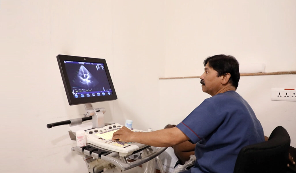
Echocardiography is a form of cardiovascular imaging that is used to obtain an understanding of the cardiac structure and function. Initially, while this assessment was being done only by 2D echo, advances has now progressed such that even 3D and 4D echocardiography is possible, for better understanding of the cardiac structure that helps in improved decision making and patient care. Both, transthoracic and transesophageal (TTE AND TEE) echocardiography is done at our OPD

Holter Monitoring 

Holter monitoring is an OPD facility, that monitors and interprets a patient’s ECG over a period of 24- 72 hours. This mainly helps in the diagnosis of abnormal heart rates and rhythms (tachyarrhythmias and bradyarrhythmias)

Hutt Test

Head up tilt test is a form of evaluation that is done for patients with syncope. It is an OPD procedure that is done under the supervision of a doctor and a nurse and assess changes in the heart rate and BP that occurs during that test, that helps to decide whether the patient will benefit from a pacemaker.

Permanent Pacemaker Implantation( Conventional And Leadless) Including Conduction System Pacing

Permanent pacemaker implantation is an inpatient procedure that is done for patients with bradyarrhythmias(with heart blocks or certain causes of syncope). Conventional form of permanent pacemaker implantation involves placement of one or two leads into the right sided cardiac chambers , followed by their connection to the pacemaker battery or pulse generator.

A new technology that has come into the market and is an approved form of therapy is leadless pacemaker implantation, that involves insertion of a small device into the right ventricle, that functions as a pacemaker. The advantage of this over the conventional pacemaker is that is reduces the complications of the same, reduces the procedure time and is easier to perform.

20-25 cases of leadless pacemaker implantation has been completed successfully at our centre, under the electrophysiology team.

Conduction Sytem Pacing

Conduction system pacing(CSP) is a new form of pacemaker implantation that aims to overcome the deficits of conventional permanent pacemaker implantation and targets the preservation of the physiological pacing pathway. CSP includes His bundle pacing and Left bundle pacing both of which are done at our centre. Close to 200 cases of conduction system pacing has been done at our centre.

arrhythmia intervention and management

Arrhythmia Intervention And Management

Tachyarrhythmias in the form of atrial fibrillation, ventricular tachycardia, AVNRT, WPW syndrome/AVRT etc, can be managed either medically or surgically. The surgical management is done in the form of EP study + RFA(electrophysiological study and radiofrequency ablation), where the abnormal electrical impulses/ pathways causing such tachyarrhythmias are mapped and these areas are ablated using radiofrequency. The EPS+RFA can be done either by 2D or 3D, both of which are available at our centre.
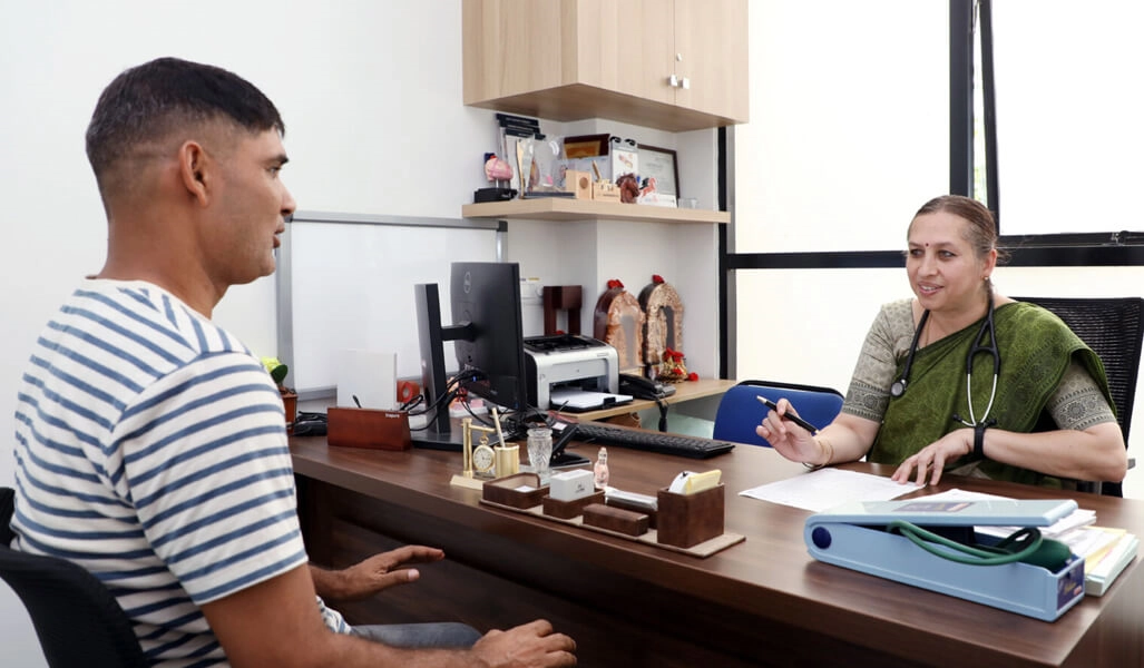
Tachyarrhythmias such as ventricular tachycardia and ventricular fibrillation can also be detected and treated on time in patients who undergo the implantation of ICD(intracardiac defibrillator) that is an invasive procedure like a pacemaker, the involves placement of a single lead in the right ventricle and its connection to a battery.

The electrophysiology unit that includes management of arrhythmias is headed by our chief electrophysiologist Dr V S Prakash.

Advanced Heart Failure Management

Heart failure can occur at different severity levels and all stages are manged successfully at our centre. Several procedures are also available to prevent worsening of initial stages of heart failure such as use of ventricular assist devices, ECMO, and CRT(Cardiac resynchronisation therapy). Cardiac transplantation, which can be a treatment modality for end stage cardiac diseases is also performed at our centre. Advanced heart failure management and cardiac transplantation is managed by our heart failure team that also includes the CTVS team and the cardiac anaesthetists

Structural Heart Disease Mangement And Intervention

Transcatheter aortic valve replacement is a recent advancement in the field of cardiology and is used as an alternative to open heart surgery and surgical aortic valve replacement. It is a minimally invasive procedure that involves the use of a catheter, inserted through the femoral artery in the leg, which is used to implant a new aortic valve. Nearly 25-30 cases of TAVR have been done successfully at our centre

Intra Coronary Imaging

A new established method that assists in the proper management of coronary artery lesions is intravascular imaging. As the name indicates, the procedure involves passing of the imaging probe into the coronary artery to give us a 360 degree view of the inside of the artery and the adjacent area and thus helps us to decide the best way to deal with the coronary artery lesions, including the need for further interventions in case of densely calcified coronary lesions, and the best size and length of the stent that is suited for the lesion. It also helps to pick up complications during or post stenting and detects inadequately opened stents.

Thus, it allows the doctor to achieve better outcomes(short and long term) of the procedure done. Intra coronary imaging is of two types- IVUS(intravascular ultrasound) and OCT( optical coherence tomography), both of which are available at our hospital

ECMO

Extracorporeal Membrane Oxygenation finds its use in multiple cardiac and non cardiac diseases. It is used mainly in critical patients to provide cardio-pulmonary support where the diseased lungs or a damaged heart are unable to support life. It pumps and oxygenates the patient’s blood outside the body and allows the patient’s heart and lungs to rest and recover

Complex Coronary Interventions And Management Of Calcified Coronary Lesions

Calcification in the walls of the coronary arteries is often a challenge to the operator and requires specific hardware and sufficient skill to deal with the same. Diagnosis of the extent of the calcium in the coronary artery and the exact site of the same can be done by intravascular imaging. Several methods are available to deal with calcified lesions, the most popular of them being, rotablation and intravascular lithotripsy(IVL), both of which are done at our centre.

Both of these methods are means of cracking/ eliminating the calcium from the walls of the coronary artery, which thus allows optimal expansion of the stent and successful angioplasty procedure

The above facilities is managed by our team of five cardiologists( Dr V K Bahl, Dr V S Prakash, Dr Anupama V Hegde, Dr Varsha R Prakash, Dr Subrahmanya Murti), two cardiac anaesthetists( Dr Guru P, Dr Nanditha), our CTVS team( Dr Ravi Shetty) and our team of perfusionists headed by Mr Sudhir.

StudyPrograms Offered

Admission 2023 group

ADDRESS

M S Ramaiah Nagar, MSRIT, Bangalore 560054